The aim of the research is to provide the eye doctors with a new tools to study the OCT images of the eye in a format intelligible for clinicians, such as quantitative maps of epiretinal abnormalities in the macular region and maps demonstrating adhesion patterns. A part the project is automatic segmentation and parameterization of retina structures such as retina layers and vessels for further support in diagnostic procedures and monitoring the progression of disorders.
Cooperation with Medical University in Poznań
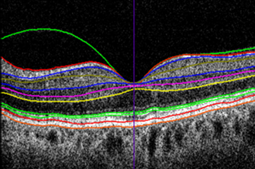
OCT B-scan with retina layers segmentations
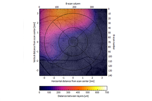
Virtual map of preretinal space volume
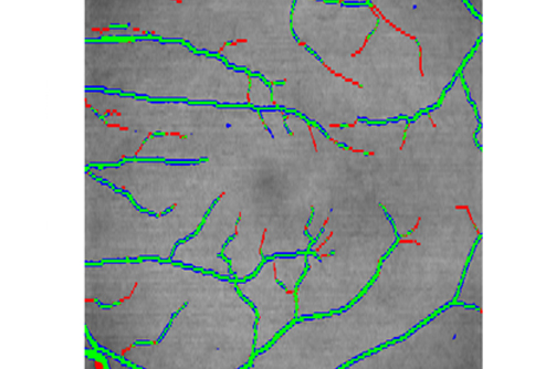
Vessel net segmentation